FIG. 4.
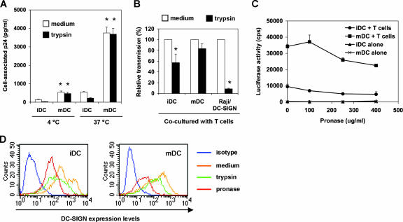
mDCs are more potent than iDCs in protecting HIV from proteolysis. (A) mDCs enhance HIV binding and internalization. DCs were incubated with HIV-Luc/JRFL (30 ng of p24) at 4°C or 37°C for 2 h, washed and treated with trypsin or medium, and then lysed for HIV p24 quantification. Asterisks indicate significant differences (P < 0.01) compared with iDCs at the same temperature. (B) DCs protect captured HIV from trypsin cleavage. HIV-pulsed iDCs, mDCs, and Raji/DC-SIGN cells were separately treated with trypsin before coculture with Hut/CCR5 target cells. Transmission of HIV-Luc/JRFL to Hut/CCR5 target cells was performed as described in the legend to Fig. 1B. The average results of four independent experiments are shown. Values for medium controls were set at 100%. Asterisks indicate significant differences (P < 0.05) between trypsin-treated samples and medium controls. (C) DCs protect captured HIV from pronase cleavage. HIV-pulsed iDCs and mDCs were treated with increasing concentrations of pronase before coculture with Hut/CCR5 target cells. The data show the means ± standard deviations of triplicate samples. One representative experiment out of two is shown. cps, counts per second. (D) Decreased surface DC-SIGN levels on DCs after protease treatment. DCs were stained for surface DC-SIGN after separate treatments with trypsin or pronase and analyzed by flow cytometry. Medium treatment was used as a control.

