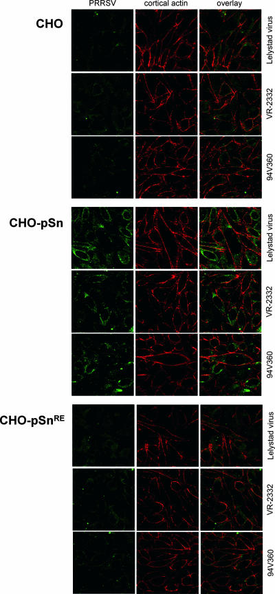
FIG. 3.
Confocal microscopic analysis of PRRSV attachment and internalization in CHO cells. PRRSV is stained green, cortical actin is stained red, and the overlay shows a superposition of the two images. Staining of the cortical actin, which lies just beneath the plasma membrane, allows discrimination of bound and internalized virus particles. The number of internalized PRRSV particles was analyzed with parental CHO cells, with CHO-pSn cells, which express recombinant porcine Sn, and with CHO-pSnRE cells, which express a mutant porcine Sn that lacks sialic acid-binding activity. Each image represents one confocal z-section through the middle of the cell.

