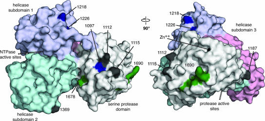
FIG. 5.
Suppressor mutations map to the surface of NS3. The surface structure of single-chain NS3-4A is shown (PDB accession code 1CU1 chain A). In this rendering, the serine protease domain of NS3 is white and NS3 helicase domains 1, 2, and 3 are purple, cyan, and pink, respectively. The cofactor peptide of NS4A (polyprotein residues 1678 to 1690) is green, and the synthetic linker between NS4A and NS3 has been removed. The locations of second- and third-site suppressor mutations are black or blue, respectively. The approximate locations of the serine protease and helicase/nucleoside triphosphatase active sites are indicated by dashed lines. This rendering was prepared with PyMOL (http://pymol.sourceforge.net/).

