FIG. 1.
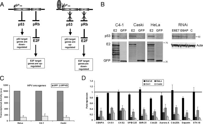
P53 stabilization and RT-PCR analysis of a selected group of mitotic genes in cervical carcinoma cell lines and HaCaT cells. (A) Schematic representation of the modulation of cellular genes through the p53 and pRb pathways in cervical carcinoma in absence or presence of the E2 transcriptional repressor. (B) Western blot analyses of the stabilization of p53 in the three cervical carcinoma cell lines expressing E2 as well as in HeLa cells transfected by the E6/E7 and the E6AP siRNA, as indicated. Expression of GFP and GFP-E2 as well as of the beta-actin is shown. (C) RT-PCR of the E6/E7 oncogenes performed in cervical carcinoma cell lines infected by adeno-GFP and adeno-GFP-E2 in three independent experiments. (D) RT-PCR of a series of cellular genes as indicated, in four keratinocyte cell lines: HaCaT not associated with HPV, HeLa and C4-1 associated with HPV18, and Caski associated with HPV16. Values given (au, arbitrary units) are levels of gene expression in adeno-GFP-E2-infected cells compared with adeno-GFP-infected cells in three independent infection experiments.

