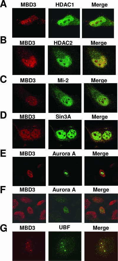
FIG. 3.
MBD3 colocalizes with UBF and Pol I in the nucleolus. HeLa cells were grown on coverslips and stained with anti-MBD3 antibody (left panel, red). The same cells were also stained with anti-HDAC1 (A), anti-HDAC2 (B), anti-Mi-2 (C), anti-Sin3A (D), anti-Aurora A (E and F, the same field with different focus levels), or anti-UBF (G) antibody (middle panel, green). Right panel, confocal merge of MBD3 with the other proteins examined.

