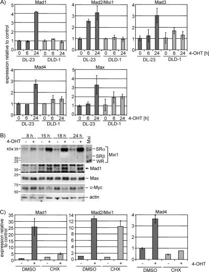
FIG. 3.
Changes in expression of members of the Myc/Max/Mxd network in response to FOXO3a activation. (A) DL23 and DLD-1 cells were treated with 100 nM 4-OHT or solvent for 6 or 24 h. Expression of Mad1, Mad2/Mxi1, Mad3, Mad4, and Max was determined by qPCR. It should be noted that CT values for Mad3 were very high, suggesting low abundance of the transcript. (B) DL23 cells were treated with 100 nM 4-OHT or solvent for 8, 10, 15, and 24 h, and total cell lysates were used to detect expression of Mxi1, Mad1, Max, and Myc by immunoblotting. Lysates from cells transfected with expression vectors for Mxi1-SRα, -SRβ, and -WR were loaded as size controls, and actin is shown as a loading control. (C) Mxi1 but not Mad1 or Mad4 is a direct target of FOXO3a. DL23 cells were stimulated with 100 nM 4-OHT in the presence of 2 μg/ml cycloheximide or dimethyl sulfoxide (DMSO) for 24 h. Expression of Mad1, Mad2/Mxi1, and Mad4 was determined by qPCR.

