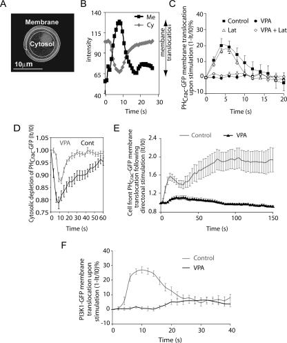
FIG. 2.
Effect of VPA on Dictyostelium PIP3 production as measured by PHCrac-GFP translocation. (A to C) Cytosolic (Cy) and membrane (Me) PHCrac-GFP (fluorescence) levels were used to monitor PIP3 production on the membrane in cells expressing PHCrac-GFP after uniform cAMP stimulation (C) with or without VPA treatment (1 mM, 10 min) or in the presence or absence of latrunculin B (Lat). (D and E) Reduction of VPA concentration (0.5 mM, 10 min) reduced and slowed the depletion of cytosolic PHCrac-GFP as a measure of PIP3 production (D), and PHCrac-GFP localization was measured at the front of chemotaxing cells in a cAMP gradient with or without VPA treatment (1 mM, 10 min) (E). (F) Cells expressing a PI3K1-GFP construct were exposed to a uniform pulse of cAMP with or without VPA (1 mM, 10 min), and PI3K-GFP membrane translocation (fluorescence) was measured at given time intervals IO and It. Data are shown as means ± standard errors (n = 8).

