
FIG. 1.
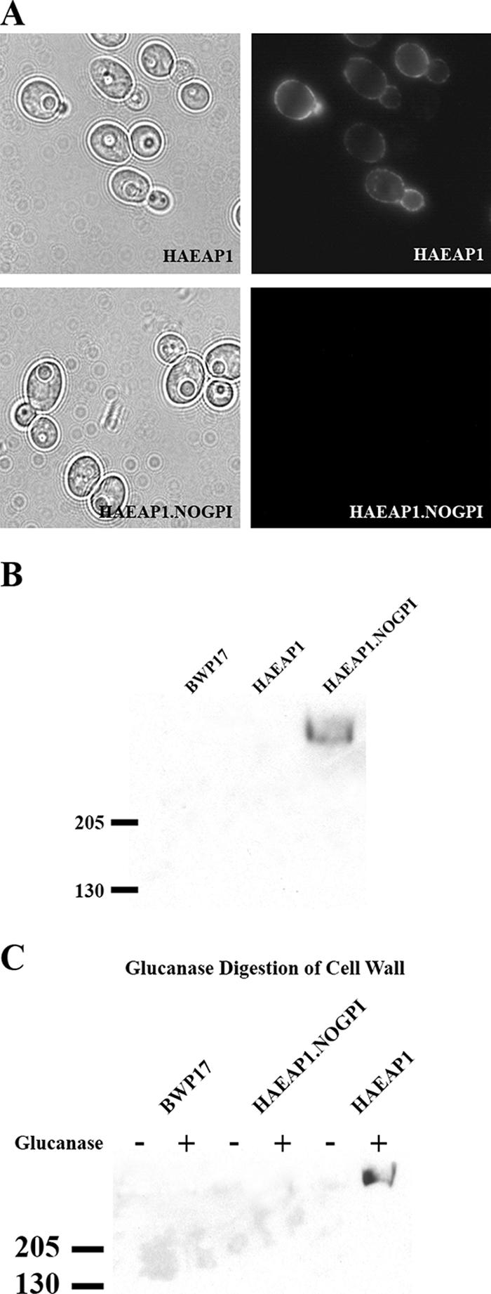
Cell wall localization of HA-tagged Eap1p. (A) Epifluorescence microscopy analysis of C. albicans strains SPY387 and SPY388. SPY387 expresses an HA-tagged Eap1p protein, and SPY388 expresses a protein identical to HA-tagged Eap1p except that 21 amino acid residues from the C terminus of Eap1p, encoding the GPI anchor signal, were deleted. Cells were cultured in SC−Ura medium overnight at 30°C prior to being imaged. (B) Analysis of cell-free supernatants from C. albicans strains BWP17, SPY387, and SPY388. The fractions were run in a 4 to 15% SDS-PAGE gradient gel and visualized by Western blotting with an anti-HA monoclonal antibody. (C) Cell wall extraction of C. albicans strains BWP17, SPY387, and SPY388. Cell walls were extracted twice in boiling SDS, digested with β-1,3-glucanase or no enzyme, and then separated into pellet and supernatant fractions. The glucanase-treated and untreated supernatant fractions were loaded into a 4 to 15% SDS-PAGE gradient gel and visualized by Western blotting with an anti-HA monoclonal antibody.