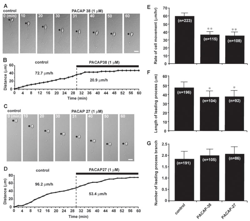
Fig. 1.

PACAP38 and PACAP27 inhibit granule cell migration in the microexplant cultures of the early postnatal mouse cerebellum. (A and C) Time-lapse images showing that application of 1 μM PACAP38 (A) or 1 μM PACAP27 (C) slows the migration of isolated granule cells in the microexplant cultures of P0–P3 mouse cerebella. Asterisks mark the granule cell somata. Elapsed time (in minutes) is indicated on top of each photograph. Scale bar: 12μm. (B and D) Sequential changes in the distance traveled by the granule cell somata shown in (A) and (C) before and afterapplication of 1 μM PACAP38 (B) or 1 μM PACAP27 (D). (E–G), Histograms presenting the changes in the rates of migration (E), the leading process length (F) and the number of the leading process branch of granule cells by application of 1 μM PACAP38 or 1 μM PACAP27. Bar: S.D. In this and following figures, single (p <0.05) and double (p <0.01) asterisks indicate statistical significance.
