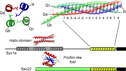
Figure 1.
Schematic representation of the different domain arrangements found in SNARE proteins. The four-helix bundle structure of the neuronal SNARE complex is shown as ribbon diagram on the top right (blue, red, and green for synaptobrevin 2, syntaxin 1a, and SNAP-25a, respectively). The layers (−7 until + 8) in the core of the bundle are indicated by virtual bonds between the corresponding Cα positions. The structure of the central 0-layer is shown in detail on the top left (Sutton et al., 1998). Below the domain architecture of two most important types of SNARE proteins are depicted. The highly conserved SNARE motif is indicated by a yellow box and the adjacent transmembrane domain by a black box. In addition, the structures of two types of autonomous N-terminal domains: a three-helix bundle (Fernandez et al., 1998; Lerman et al., 2000) and a profilin-like domain (Gonzalez et al., 2001) are shown.

