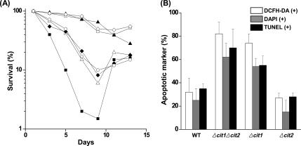
Figure 5.
Deletion of CIT1 causes accelerated aging associated with typical hallmarks of apoptosis and subsequent adaptive regrowth. (A) Analysis of the rate of aging of yeast strains. Yeast strains (wild type, Δcit1, Δcit2, Δcit1Δcit2, Δcit1Δcit2/YEpCit1, Δcit1Δcit2/YEpCit2, and Δcit1/YEpCit2) were grown in SCD for 13 d. Aliquots of the cultures were taken at intervals, and the portion of living cells was then monitored by CFU assays carried out in triplicate. □, wild type; ▵, Δcit1; ▴, Δcit2; ■, Δcit1Δcit2; ◇, Δcit1Δcit2/YEpCit1; ○, Δcit1Δcit2/YEpCit2; ♦, Δcit1/YEpCit2. (B) Percentage of cells exhibiting the typical hallmarks of apoptosis in aged cultures. The yeast strains listed above were grown in SCD for 7 d. To estimate the proportion of ROS-accumulating cells, yeast cells were stained with DCFH-DA, and then they were examined by fluorescence microscopy. DCFH-DA–positive cells were counted in fluorescence images and total cells in corresponding DIC images. For observation of nuclear fragmentation, cells were stained with DAPI, and the proportions of the cells with abnormal nuclei were estimated by fluorescence microscopy. For observation of DNA fragmentation, cells were stained with TUNEL reagent. The proportions of cells exhibiting DNA fragmentation were estimated by bright-field microscopy.

