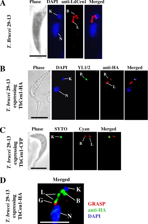
Figure 7.
(A) Immunolocalization of TbCen1 in the procyclics of T. brucei 29-13 using anti-LdCen1 Ab (stains LdCen1 at the basal body in L. donovani (Selvapandiyan et al., 2004) stains here the basal body and the bilobe. (B) Localization of TbCen1-HA proteins in T. brucei 29-13 procyclic cells transformed with pLEW100-TbCen1. Staining with anti-HA Ab stains the basal body and the bilobe, and YL1/2 stains the basal body. (C) Fluorescent images of a live T. brucei cell ectopically expressing TbCen-CFP are stained with cyan, and kinetoplast was stained with SYTO Green-Fluorescent Nucleic Acid Stain (Molecular Probes, Invitrogen). (D) Localization of TbCen1 with respect to Golgi. Cells were stained with GRASP to stain the Golgi and anti-HA Ab to stain TbCen1-HA at the basal body and the bilobe. In all, the parasites obtained were from midlog culture and also stained with DAPI for the nuclei and the kinetoplasts. K, kinetoplast; N, nucleus; B, basal body; G, Golgi; L, bilobe. Scale bar in all, 5 μm.

