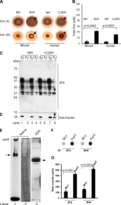
Figure 1.
(A) Equal aliquots (5 μl each) of 10% brain homogenate from normal human and mouse (NH), scrapie-infected mouse (ScH), and sCJD were blotted on PVDF membrane and reacted for ferrous and ferric iron (Smith et al., 1997). In contrast to NH, ScH and CJDH show strong reactivity for ferrous iron [Fe(II)]. Ferric iron is detected in all samples, but significantly more is in ScH and CJDH samples [Fe(III)]. (B) Colorimetric quantification of total iron shows four- to sixfold more iron in ScH and CJDH samples compared with age-matched NH controls. Unpaired t test shows highly significant mean difference between normal and diseased samples. Values for mouse samples: Δmean = 36.7; 95% confidence interval (CI) = 44.3, 29.2; t = 13.5; df = 8; p < 0.0002. Values for human samples: Δmean = 25.5; 95% CI = 29.2, 21.9; t = 19.6; df = 4; p < 0.0001). (C) Lysates of PrPC cells exposed to biotin-tagged NH or CJDH were subjected to differential centrifugation and immunoblotted with 3F4. In NH-exposed lysates, most of PrPC fractionates in the detergent-soluble S1I and S2I fractions (lanes 1 and 3). Minimal amounts of unglycosylated PrPC are detected in the low- and high-speed detergent-insoluble fractions P1I and P2I (lanes 2 and 4). In contrast, CJDH-treated lysates show a significant amount of PrP in the high-speed detergent-insoluble P2I fraction (lanes 5–8). (D) Reprobing with anti-ferritin shows slight up-regulation of ferritin in CJDH-treated lysates, 40% of which partitions in the P2I fraction (lanes 1–8, arrow). (E) Recombinant PrP radiolabeled with 59FeCl3–acetate complex shows a prominent iron-labeled band on native iron gel (lane 1) that shows a negative stain when stained with silver due to bound iron (lane 2). Minor bands that stain with silver probably represent degradation products that do not bind iron. Fractionation on SDS-PAGE and silver staining shows a single prominent band of recombinant PrP (lane 3). (F) M17 and PrPC cells were radiolabeled with 59FeCl3–citrate complex for 4 h and subjected to immunoprecipitation with 3F4 and 8H4. Eluted proteins were spotted on PVDF membrane followed by autoradiography. (G) Quantification of iron-labeled proteins in the immunoprecipitate by direct gamma-counting. n = 5. Unpaired t test shows highly significant mean difference between iron-labeled PrP immunoprecipitated from M17 and PrPC cells. Values for 3F4 immunoprecipitates: Δmean = 313.1; 95% CI = 348.9, 277.3; t = 24.3; df = 8; p < 0.0001. Values for 8H4 immunoprecipitates: Δmean = 362.5; 95% CI = 404.2, 320.8; t = 24.15; df = 8; p < 0.0001.

