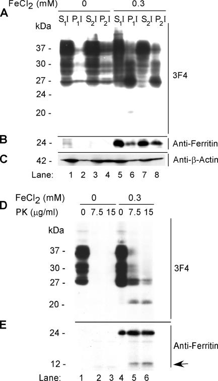
Figure 3.
(A) Mock- and 0.3 mM FeCl2-exposed cell lysates were fractionated by differential centrifugation and immunoblotted with 3F4. As expected, the majority of PrPC from mock-treated lysates partitions in the detergent-soluble S1I and S2I fractions (lanes 1 and 3), with minimal amounts in the P1I and P2I fractions (lanes 2 and 4). In contrast, significant amounts of PrPC from FeCl2-treated lysates are detected in the pellet fractions P1I and P2I (lanes 6 and 8). A minor 20-kDa fragment is observed in the P1I fraction of treated cells (lane 6). (B) Reprobing with anti-ferritin shows up-regulation of ferritin in FeCl2-treated lysates (lanes 1–8), 40% of which partitions in the P2I fraction (lane 8). (C) FeCl2 treatment has no detectable effect on the expression or solubility of β-actin. (D) Treatment of lysates prepared from mock- and FeCl2-exposed cells with 7.5 and 15 μg/ml PK results in the complete digestion of PrPC in mock-treated controls (lanes 1–3), whereas treated lysates show a 20-kDa PK-resistant fragment (lanes 4–6). (E) Reprobing with anti-ferritin shows up-regulation of ferritin in FeCl2-exposed cells (lanes 1–6) and the generation of a 14-kDa fragment by PK (lanes 5 and 6, arrow).

