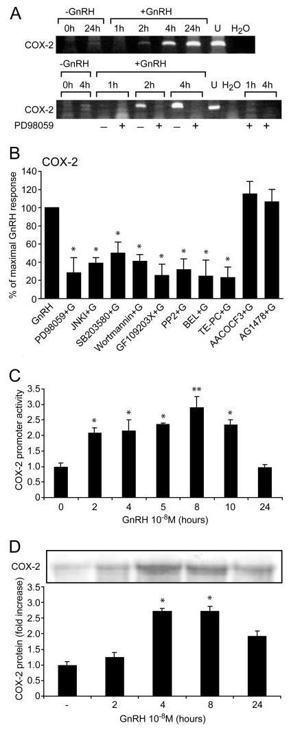
Fig. 1. GnRH Induction of COX-2 Activity.

A, Effect of GnRH on COX-2 induction as revealed by RT-PCR. Subconfluent LβT2 cells were incubated and some groups were pretreated with the selective MEK inhibitor PD98059 (50 μm for 20 min) before the addition of GnRH (10 nm) for the time indicted. COX-2 was then determined by qualitative RT-PCR. Negative (H2O) and positive (U, uterus) controls are also shown. B, Quantitative-PCR for COX-2 induction by GnRH. Subconfluent LβT2 cells were pretreated for 20 min with the following selective inhibitors: for MEK (PD98059, 50 μm), for JNK (JNK inhibitory peptide, 2 μm), for p38 (SB203580, 10 μm), for PI3K (Wortmannin, 25 nm), for PKC (GF109203X, 3 μm), for c-Src (PP2, 5 μm), for iPLA2 (BEL, 20 μm), for sPLA2 (TE-PC, 25 μm), for cPLA2 (AACOCF3, 25 μm) and for EGF receptor kinase (AG1478, 5 μm). Cells were then stimulated with GnRH (100 nm for 8 h) and COX-2 was determined by Q-PCR. C, Effect of GnRH on COX-2 promoter activity. Subconfluent LβT2 cells were seeded into six-well plates and incubated overnight at 37 C before transfecting with 0.3 μg of −2307/+49 human COX-2 promoter. Renilla (33 ng) expression vector was also included as a measure of transfection efficiency and as an internal control. The transfected cells were serum starved for 18 h. GnRH (100 nm) was added for the indicated time points and cells were then harvested and assayed using a Dual-light Luciferase assay kit (Promega) in a FLUOStar Optima luminometer. Luciferase activity was normalized for Renilla to correct for transfection efficiency. Results for promoter activity are expressed as fold increase relative to untreated controls (n = 6). D, Effect of GnRH on COX-2 protein expression. LβT2 cells were grown in 4 × 12-well plates (5 × 10⁵ cells/well). Cells were incubated in serum-free DMEM, 0.2% FCS overnight at 37 C. The cells were washed with DMEM and incubated with or without GnRH (100 nm) for various time periods and COX-2 was detected by Western blotting. The blot was re-run with antitotal ERK antibody to correct for equal loading of the samples. A representative gel is shown, and bars are the mean from triplicate samples (ANOVA: *, P < 0.05, **, P < 0.01 compared with control).
