
Figure 4.
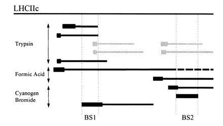
[14C]DCCD-labeled fragments of LHCIIc apoprotein. Labeled fragments of LHCIIc (solid bars) generated by cleavage with trypsin, formic acid, or CNBr were identified by estimation of their molecular weights (from their electrophoretic mobility), and comparison of their N-terminal amino acid sequences (thick bars) with the consensus sequence for LHCIIc apoprotein. Their positions are shown relative to the full LHCIIc apoprotein. In two cases, amino acid sequence data were ambiguous due to the presence of two comigrating fragments (gray bars). The regions of overlap identifying DCCD-binding sites (BS1, BS2) are bordered by dotted vertical lines, and begin at the Met residues identified in Fig. 3.