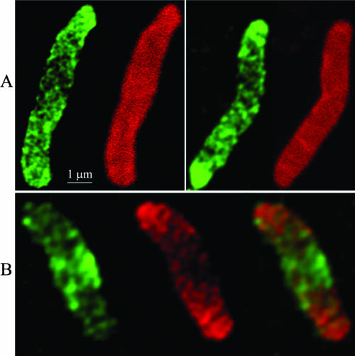
FIG. 5.
Inhibiting MreB alters the pattern of peptidoglycan insertion into the cell wall. AV23-1 was filamented by inhibiting FtsZ with SulA expressed from pFAD38, the cells were labeled with d-cysteine for 90 min and chased for 30 min at 42°C in the presence of A22 (5 μg/ml) and in the absence of d-cysteine, and sacculi were prepared and immunolabeled. (A) d-Cysteine pulse-chase-labeled sacculi. The left-hand cell in each panel displays older peptidoglycan (green) with uneven, localized incorporation of new material (dark areas). The right-hand cell in each panel is immunostained to detect all peptidoglycan; uniform labeling indicates that the dark areas are not the result of murein degradation. (B) Vancomycin labeling of a typical d-cysteine-labeled sacculus. The left-hand figure shows the location of vancomycin-labeled peptidoglycan (green), the middle figure shows the location of older d-cysteine-labeled peptidoglycan (red) and the right-most figure is a merged image. Note that the location of vancomycin-labeled material corresponds to the d-cysteine-free central dark area.

