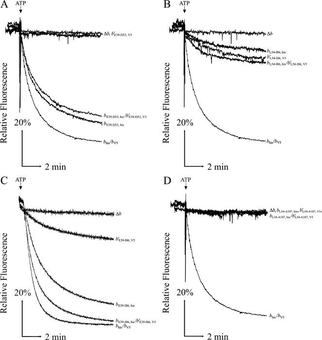
FIG. 3.
ATP-driven energization of membrane vesicles prepared from KM2 (Δb) cells expressing chimeric b subunits. Aliquots of membrane proteins (500 μg) were suspended in 3.5 ml assay buffer (50 mM MOPS [pH 7.3], 10 mM MgCl2), and fluorescence quenching of ACMA was used to detect energization of membrane vesicles after the addition of ATP as previously described (33). Traces are plotted as relative fluorescence over time. The point where ATP was added is indicated above each graph, and chimeric subunits are given on the right. Each panel shows traces of membranes from the negative control KM2/pBR322 (Δb), the positive control KM2/pTAM37/pTAM46 (bhis/bV5), and the b and b′ chimeric subunits expressed individually and together. The chimeric subunits in the panels are as follows: E39 to D53 (A), L54 to I86 (B), E39 to I86 (C), and L54 to A107 (D). Traces were obtained using a Perkin-Elmer (A, B, and D) or Photon Technologies International (C) spectrofluorometer.

