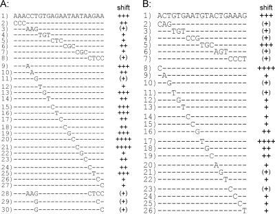
FIG. 6.
Mutational analysis of the PhoR-binding sites within the pstS promoter (A) and the phoR promoter (B). Previous experiments had indicated that fragments 1 have a high affinity to PhoR∼P. To determine the relevance of individual base pairs for binding, fragments with different mutations were tested in gel mobility shift assays for the ability to bind PhoR∼P (for details, see the legend to Fig. 3). According to the results of the shift (see Fig. S1 in the supplemental material), the fragments were divided into five categories: ++++, 50% of the fragment shifted with a <30-fold molar excess of PhoR∼P, +++, 50% of the fragment shifted with an ∼30-fold molar excess of PhoR∼P; ++, 50% of the fragment shifted with a >30- to 45-fold molar excess of PhoR∼P and 100% of the fragment shifted at a 60-fold molar excess of PhoR∼P; +, <50% of the fragment shifted with a >30- to 45-fold molar excess of PhoR∼P and 100% shifted at a 90-fold molar excess of PhoR∼P; and (+), 50% of the fragment shifted at a >45- to 90-fold molar excess of PhoR∼P.

