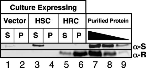
FIG. 4.
Western blots comparing in vivo levels of expression of His6-RhaS-CTD and His6-RhaR-CTD. Soluble (S, supernatant) and insoluble (P, pellet) fractions after sonication were loaded as indicated. The vector-only sample was pSU18. His6-RhaS-CTD (HSC) was expressed from pSE271, and His6-RhaR-CTD (HRC) was expressed from pSE272. Lanes 7 to 9 on each gel contained known amounts of purified His6-RhaS-CTD (top blot) or His6-RhaR-CTD (bottom blot). The amounts of His6-RhaS-CTD were 737 (lane 7), 368 (lane 8), and 184 (lane 9) ng. The amounts of His6-RhaR-CTD were 162 (lane 7), 54 (lane 8), and 18 (lane 9) ng. Two gels were prepared, with identical culture samples on each gel, and each blot was probed with the primary antibody corresponding to the purified protein samples loaded, as indicated to the right. α-S, anti-RhaS antibody; α-R, anti-RhaR antibody.

