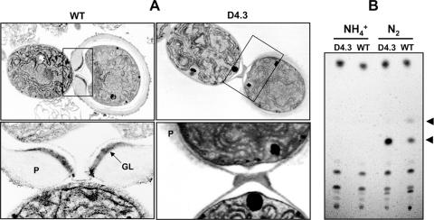
FIG. 5.
The ultrastructure of heterocysts (A) and components of heterocyst glycolipids (B). (A) Electron microscopic images of the wild type (WT) and strain D4.3. Filaments were cultured in the absence of a fixed-nitrogen source. Boxed areas in the upper panels were enlarged with higher magnification in the lower panels. P, polysaccharide layer; GL, glycolipid layer. (B) Thin-layer chromatographic analysis of glycolipids in the wild type (WT) and D4.3. Cells were cultured in the presence of ammonium (NH4+) or in the absence of combined nitrogen (N2). The heterocyst-specific glycolipids are indicated by arrows; the upper one (the minor HGL) corresponds to 1-(O-α-d-glucopyranosyl)-3-keto-25-hexacosanol, and the lower one (the major HGL) corresponds to 1-(O-α-d-glucopyranosyl)-3,25-hexacosanediol.

