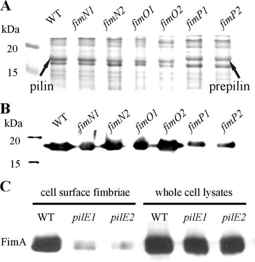
FIG. 3.
Western immunoblotting of whole-cell lysates and purified cell surface fimbriae. Shown is the production of FimA in whole-cell lysates from the wild-type strain VCS1703A (WT) and the mutants JIR3895 (fimN1), JIR3896 (fimN2), JIR3885 (fimO1), JIR3886 (fimO2), JIR3889 (fimP1), and JIR3890 (fimP2). Samples were separated by 12 to 15% SDS-PAGE and analyzed by Coomassie brilliant blue staining (A) and Western immunoblotting with fimbrial serogroup G-specific antisera at a 1:1,000 dilution (B). Note that the FimA-specific protein is larger in the fimP mutants. (C) Reduced extracellular fimbrial levels of pilE mutants. Purified cell surface fimbriae and whole-cell lysates of wild-type strain VCS1703A (WT) and the mutants JIR3910 (pilE1) and JIR3911 (pilE2) were analyzed.

