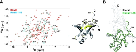
FIG. 5.
Superposition of full-length RimM and RimM.1-85. (A) Left, 1H-15N HSQC spectra of full-length RimM (red) and RimM.1-85 (blue), recorded at 25°C on a 600-MHz spectrometer. Most of the resonances originating from the N-terminal domain were overlapped with each other. Only the shifted resonances for residues 1 to 85 of full-length RimM, compared to those for RimM.1-85 itself, are labeled with the residue number and the one-letter amino acid code: V4, E5, I6, G7, A14, R22, E24, V26, V27, H29, L30, E31, R32, G71, D80, L81, and E85. Right, mapping of these residues (yellow) on the RimM.1-85 structure. (B) Superposition of the backbones of the 10 best structures of full-length RimM (black) and the best structure of RimM.1-85 (green). The 10 structures of full-length RimM were calculated with CYANA 1.0.8. The structure of the C-terminal region (residues 96 to 162) is not displayed, because it does not adopt a rigid tertiary structure.

