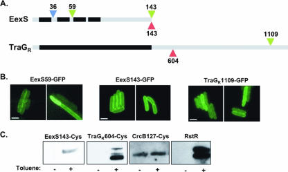
FIG. 2.
TraGR and EexS exclusion residues are localized in the cytoplasm. (A) Schematic depiction of the EexS and TraGR proteins with the predicted transmembrane (black) and soluble (gray) regions. Residue numbers and colors indicate the sites where GFP (green), PhoA (blue), and cysteine (red) insertions were placed. (B) Fluorescence images of cells expressing the indicated GFP fusion constructs. Bars represent 2 μM scales. (C) Detection of biotinylation (see the discussion of methods in the supplemental material) of the indicated proteins from cells either left untreated or treated with toluene to permeabilize the membrane. Estimated protein sizes are as follows: EexS143-Cys, 18 kDa; TraGR604-Cys, 128 kDa; CrcB127-Cys, 13 kDa; RstR, 10 kDa. Residue 127 of CrcB is known to be periplasmic, while RstR is a known cytoplasmic protein.

