FIG. 5.
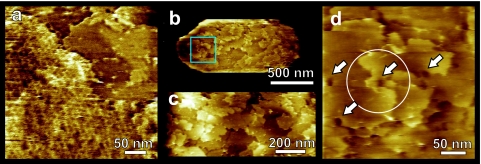
C. novyi NT spore coats: high-resolution AFM height images. Spores were activated and exposed to germination medium as described in the text. (a) Most of the honeycomb layers disappeared from the spores within ∼1 h. Remaining honeycomb patches (left, lower sides) could be easily removed by scanning with increased force. Below the honeycomb layer several underlying coat layers (upper right) are revealed. (b to d) Typical growth patterns seen on C. novyi NT spore surface after removing the honeycomb layers. (b) Whole spore with several ∼6-nm-thick layers exposed on the surface. (c) Zoom-in of the center of panel b showing that spore coat layers originate at screw dislocations. (d) Zoom-in of the area indicated in panel b. The circle in panel d denotes a fourfold screw axis. Many dislocation centers show depressions reminiscent of hollow cores (arrows), which are found in a wide range of crystals. The times in germination medium (hours:minutes) were 5:40 (a) and 4:10 (b to d).

