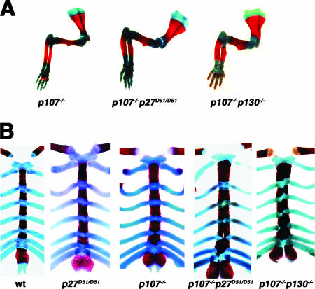
FIG. 3.
Endochondral ossification in newborn mice. Neonatal mice were collected, and skeletal preparations were stained with alcian blue and alizarin red to differentiate the chondrocytic and bony regions. (A) Representative images of the forelimbs from p107−/−, p107−/− p27D51/D51, and p107−/− p130−/− mice are shown. Red, bone; blue, cartilage. The number of animals analyzed for this phenotype is indicated in Table 3. (B) Representative images of the sternal regions of mice. Note the presence of bone in the costal junctions in p107−/− p130−/− mice and the p107−/− p27D51/D51 deficient mice. The number of mice afflicted with this phenotype is reported in the text.

