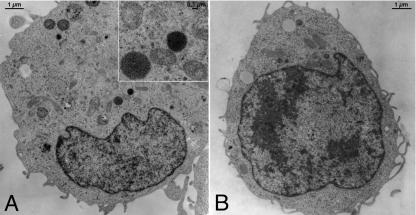
FIG. 2.
Electron microscopy of a semiadherent HMC-1 cell without (A) and with (B) treatment with TcdB (3 nM) for 90 min. Typical mast cell-specific granules (granules filled with electron-dense [black] material; inset in panel A) are present in these cells, which were presumably exocytosed after stimulation by TcdB.

