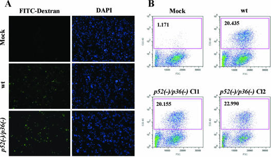
FIG. 2.
p52/p36-deficient sporozoites traverse and wound cells normally. (A) Overview of infected HepG2-CD81 cultures showing FITC-positive (green) wounded cells as a result of wt or p52/p36-deficient sporozoite traversal. DAPI (blue) visualizes the nuclei. (B) Quantification of cell-wounding activity using flow cytometry reveals a similar level of traversal activity in wt and p52/p36-deficient sporozoites. The assay was repeated six times for each sample. Approximately 3.0 × 10⁴ sporozoites were incubated with 5.0 × 10⁴ subconfluent HepG2-CD81 cells per well in the presence of FITC-dextran. The x axis represents the forward-scatter properties of the cells, while the y axis represents the green fluorescence. The numbers of wounded cells (percent) are shown in the upper left corners of the graphs. Approximately 20% of the HepG2-CD81 cells inoculated with wt and p52/p36-deficient (Cl1 and Cl2) sporozoites were fluorescent, i.e., wounded. Mock infections were done by incubating HepG2-CD81 cells with uninfected mosquito salivary gland preparations.

