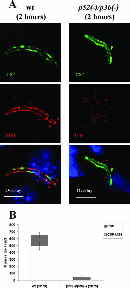
FIG. 5.
p52/p36-deficient parasites fail to establish infection in the host liver. (A) Indirect immunofluorescence assay of wt and p52/p36-deficient parasites detected in the host liver at 2 h p.i. The upper panels show intracellular wt parasite or p52/p36-deficient parasite staining with anti-CSP Abs. The PVM is visualized in wt parasites using anti-UIS4 Ab staining, but UIS4 staining is not discernable in p52/p36-deficient parasites, indicating a PVM deficiency (middle panels). The overlay of UIS4 staining, CSP staining, and nuclear DAPI staining is shown in the bottom panels (scale bar, 20 μm). (B) Quantification of liver infections. Numbers shown are means ± standard deviations of wt parasites and p52/p36-deficient parasites detected in three discontinuous sections of livers of BALB/c mice 2 h p.i. p52/p36-deficient parasites were detected at greatly reduced numbers (∼90% reduction) compared to wt parasites and did not show UIS4 staining (P < 0.0001, Fisher's exact test). Approximately 75% of wt parasites detected at 2 h p.i. showed UIS4-positive staining and CSP-positive staining.

