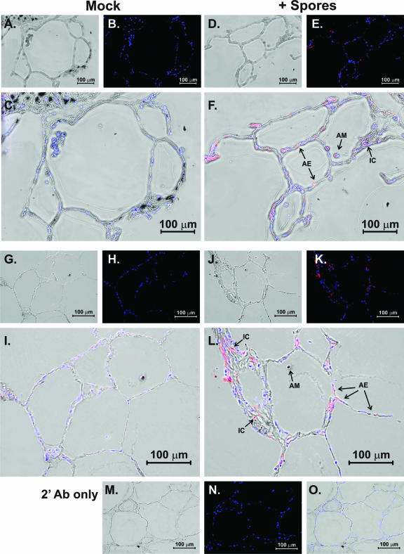
FIG. 6.
Cellular source of cytokine and chemokine induction by B. anthracis spores. Lung slices were exposed to 1 × 10⁶ spores or spore diluent for 24 h in the presence of brefeldin A to enhance detection of cytokines. The slices were then processed for immunohistochemistry analysis for detection of the cytokine IL-6 (A to F) and the chemokine IL-8 (G to L) using goat polyclonal antibodies as described in Materials and Methods. Panels A, D, G, J, and M are bright-field images that demonstrate that lung architecture was preserved during the experiment. Panels B, E, H, and K are fluorescent images that show staining of nuclei by DAPI (blue) and IL-6 (B and E) or IL-8 (H and K) detection by Alexa Fluor 546 (red) secondary antibody. Panels C, F, I, L are overlays of the bright-field and fluorescent images and demonstrate that the primary cellular sources of the cytokines are alveolar epithelial cells (AE) and alveolar macrophages (AM). Some interstitial cells (IC) are also positive for IL-6 or IL-8. Panels M, N, and O are bright-field, fluorescent, and overlaid images of spore-exposed tissue stained with only secondary antibody (2′ Ab) and confirm that omission of the primary antibody results in a loss of signal.

