FIG. 8.
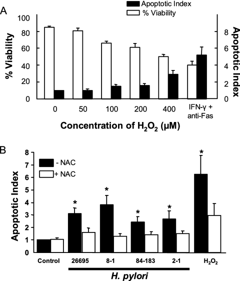
Induction of apoptosis in gastric epithelial cells and its inhibition by antioxidants. (A) NCI-N87 cells were exposed to H2O2 at concentrations from 0 to 400 μM with cell viability (expressed as % of control) measured by trypan blue exclusion and apoptosis (shown as apoptotic index), determined by detection of endonucleosomes via ELISA. Cells exposed to 800 U/ml IFN-γ for 6 h followed by 100 μg/ml anti-Fas antibody were used as a positive control. Dose-dependent changes were observed with decreases in cell viability and increases in apoptosis significantly different from those of control cells at doses of 100 to 400 μM H2O2 (P < 0.05). Data shown as means ± SEM (n = 3 to 5). (B) Kato III cells were treated with H. pylori (300 bacteria per epithelial cell), 400 μM H2O2, or media alone (control) for 48 h in the presence or absence of 10 mM NAC. Apoptosis was assessed using an ELISA to detect endonucleosomes exposed by DNA fragmentation. Values depicted are means ± SEM (n = 9 to 15). *, P < 0.05 compared to cells without NAC.

