FIG. 1.
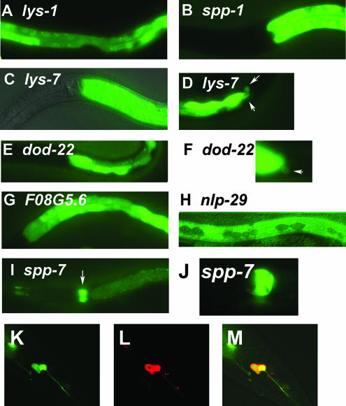
C. elegans candidate antimicrobial genes are expressed in tissues exposed to the environment. Panels A to J depict representative fluorescence micrographs of the indicated GFP fusion-bearing strains (panels C and H are overlays of fluorescence and Nomarski micrographs). Strong intestinal gfp expression is observed in panels A (mid-body view), B, C, E, and G (anterior to mid-body view), and D and F (posterior view). Weak intestinal gfp expression and strong pharyngeal gfp expression are observed in panel I. Panel J is a close-up view of the pharyngeal expression in panel I. White arrows point to rectal gland gfp expression in panels D and F or to pharyngeal expression in panel I. Panels K, L, and M are confocal microscopy images of the posterior part of the same nematode expressing lys-1::gfp. Panel K depicts gfp expression (green), panel L depicts animals filled with the dye DiI, which labels some chemosensory neurons (red), and panel M depicts the overlap (yellow), demonstrating that this gfp fusion is expressed in two chemosensory phasmid neurons. Anterior is to the left and ventral is down in all images. A complete summary of the gfp expression data for all 14 antimicrobial::gfp fusions is presented in Table 1.

