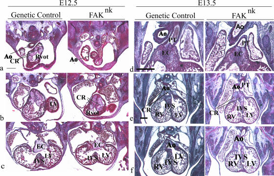
FIG. 4.
FAK is essential for normal OFT cushion morphogenesis. Mason's trichrome or hematoxylin-eosin-stained sections from control and FAKnk embryos at E12.5 and E13.5. (a) At E12.5, the aortic OFT was anterior to and to the right of the right ventricular OFT in the FAKnk hearts. (b) The endocardial cushions appear normal in the OFT in the FAKnk hearts at E12.5. (c) The AV canal endocardial cushions, the muscular IVS, and the trabeculae appear normal in the FAKnk hearts (right) compared to genetic controls (left) at E12.5. (d) At E13.5, the conal ridges had proliferated and fused along the midline in the distal OFT in the genetic control (left) and FAKnk (right) hearts. (e) At E13.5, the conal ridges had fused normally in the proximal OFT with the muscular IVS in the genetic control hearts (left), but this fusion was abnormal in the FAKnk hearts (right). (f) At E13.5, the aorta maintained contact with the left ventricle in the genetic control hearts (left), but in the FAKnk hearts, the aorta maintained contact with the right ventricle (right). Ao, aorta; CR, conal ridges; EC, endocardial cushion; LV, left ventricle; mV, mitral valve; PT, pulmonary trunk; Rvot, right ventricular OFT; RV, right ventricle; tv, tricuspid valve. Scale bars, 100 μm.

