FIG. 7.
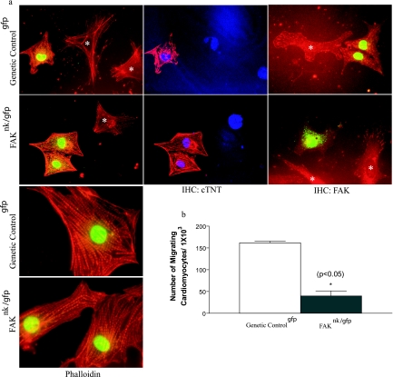
Depletion of FAK does not affect myofibrillar organization but impairs cardiomyocyte motility. (a) Cells were isolated from genetic controlgfp and FAKnk/gfp hearts and processed for immunohistochemistry (IHC) as described in Materials and Methods. GFP-positive cells from the genetic controlgfp and FAKnk/gfp hearts showed a characteristic striated pattern of sarcomeric actin, as revealed by phalloidin staining (left), and high levels of cTNT (middle), as determined by immunohistochemistry. GFP-negative cells (*) are likely cardiac fibroblasts. Cells were also stained with anti-FAK antibodies to reveal the presence or absence of FAK in both myocytes and nonmyocytes (*) isolated from the genetic controlgfp and FAKnk/gfp hearts (right). (b) GFP-positive cells isolated from genetic controlgfp and FAKnk/gfp hearts were plated on Boyden chambers coated with fibronectin (10 μg/ml) as described in Materials and Methods. The data are presented as the mean number of GFP-positive migrating cells compared to the total of GFP-positive attached cells (± standard deviation) counted prior to addition of the chemoattractant (15% serum). The data are representative of cardiomyocytes isolated from three separate hearts from each genotype.

