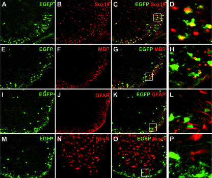
FIG. 5.
Cell-type-specific expression of the MBP-Sox4 transgene in the spinal cord. Antibodies directed against EGFP (in green) (A, E, I, and M) were combined with antibodies directed against the oligodendroglial marker Sox10 (B), the marker for differentiating oligodendrocytes, MBP (F), the astrocyte marker GFAP (J), and the panneuronal marker NeuN (N) (all in red) in coimmunohistochemistry (C, D, G, H, K, L, O, and P) on sections of transgenic spinal cord at postnatal day 3 (A to H) and postnatal day 7 (I to P). Coexpression of EGFP and cell-type-specific marker is visible as a yellow signal in panels C, D, G, H, K, L, O, and P. Panels D, H, L, and P represent magnifications of the boxed area in panels C, G, K, and O.

