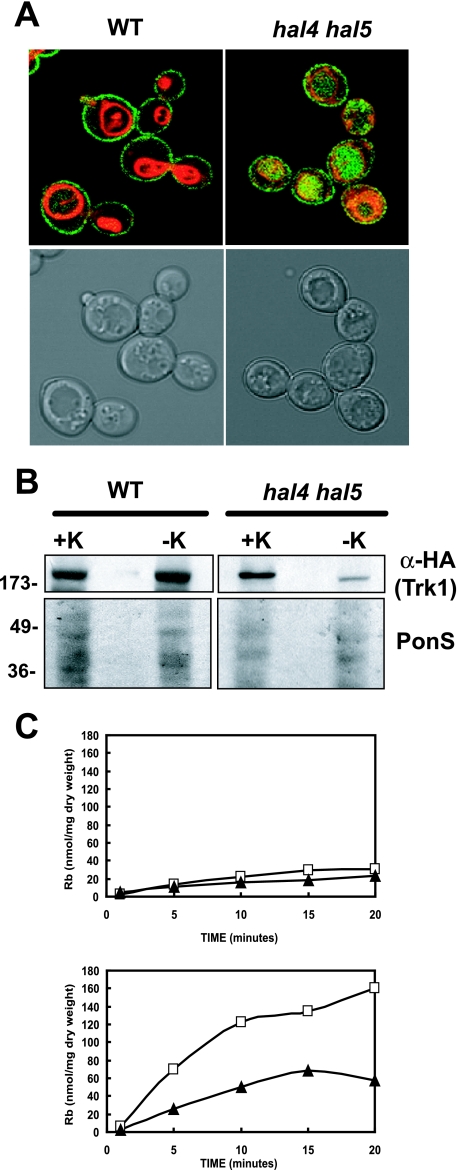
FIG. 1.
Trk1 is less stable in the hal4 hal5 mutant upon potassium starvation. (A) The wild-type (WT) strain and the hal4 hal5 mutant expressing a centromeric plasmid containing a TRK1-GFP fusion were grown to mid-log phase in potassium-supplemented medium and stained with the vacuolar dye FM4-64 as described in Materials and Methods. Gray scale and fluorescence overlays of representative confocal microscopy images depicting the localization of Trk1 (green) and the vacuolar membrane (red) are shown. (B) Western analysis of Trk1 levels in the membrane fractions of the wild-type and hal4 hal5 mutant strains maintained in potassium chloride-containing minimal medium (+K) or incubated in unsupplemented medium for 2 h (−K). Images of the Ponceau S-stained filters are shown in the bottom panels, as a control for protein loading. (C) The wild-type (□) and hal4 hal5 (▴) strains were grown to mid-log phase in minimal medium supplemented with potassium chloride and were analyzed for high affinity rubidium uptake (0.5 mM) immediately after washing (top panel) and after 2 hours of incubation in low-potassium medium (bottom panel). Similar results were observed using 50 mM RbCl.

