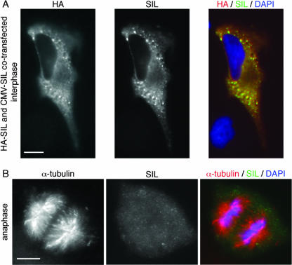
FIG. 6.
SIL is expressed in the cytoplasm when overexpressed, and SIL does not localize to the spindle poles during anaphase. (A) HeLa cells were cotransfected with pCMV-HA-SIL and pCMV-SIL and immunostained with anti-HA (red) and anti-SIL (green) antibodies. SIL was present in the cytoplasm and stains positively with both antibodies. As in the cell shown, high levels of overexpression resulted in the formation of positively stained foci, which may be accumulation of proteins at the ribosomes or protein aggregates. CMV, cytomegalovirus. (B) A cell in anaphase stained with the anti-SIL antibody reveals that SIL does not localize to the spindle poles when cells are in anaphase. Scale bars = 10 μm.

