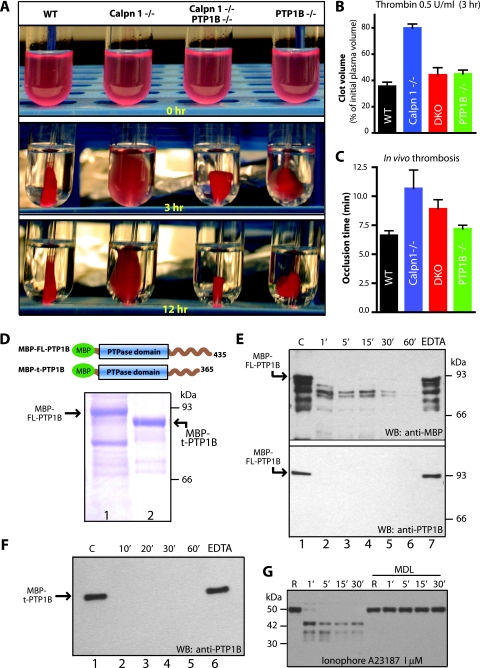
FIG. 7.
Correction of clot retraction defect in the double knockout mice. Platelet-rich plasma samples were pooled, and platelet counts were normalized. (A) Representative photographs of clot retraction induced by thrombin. (B) Quantification of fibrin clot retraction 3.0 h after thrombin and CaCl2 treatment. Clot volumes are expressed as a percentage of initial platelet-rich plasma volume. (C) Measurement of in vivo thrombosis by ferric chloride injury assay. Calpain-1 null mice are less susceptible to thrombus development. The mean plus SEM (error bar) for each group are shown. Data were analyzed using one-way analysis of variance by GraphPad prism software (P = 0.01). (D) Coomassie blue staining of recombinant PTP1B. Bacterially expressed full-length PTP1B (FL-PTP1B) (lane 1) and truncated PTP1B (t-PTP1B) (lane 2) were purified by affinity chromatography. The boundaries of each fusion protein with MBP at the N terminus are shown in the schematic diagram. (E) In vitro proteolysis of full-length recombinant PTP1B (FL-PTP1B) by commercial calpain-1 for indicated time intervals (from 1 min [1′] to 60 min [60′]) (see Materials and Methods for details). Lane 1 contains a negative control where calpain-1 was not added. Lanes 2 to 6 show various incubation times of PTP1B with calpain-1. Lane 7 shows incubation of PTP1B with calpain-1 in the presence of 2.0 mM EDTA for 60 min to inhibit calpain-1. The top blot was probed with a rabbit polyclonal antibody raised against MBP. The bottom blot was probed with a commercially available polyclonal antibody against PTP1B. WB, Western blotting. (F) In vitro proteolysis of truncated PTP1B by calpain-1. Lane 1 contains a negative control with no calpain-1, and lanes 2 to 5 show incubation times (from 10 min [10′] to 60 min [60′]) with calpain-1. Lane 6 shows incubation with calpain-1 in the presence of 2.0 mM EDTA for 60 min. Western blotting (WB) was carried out using an anti-PTP1B antibody. (G) Gel-filtered platelets (3 × 10⁸/ml) from WT mice were incubated with either DMSO or calpain inhibitor MDL 28170 (350 μM) in DMSO for 30 min at room temperature. PTP1B proteolysis was initiated by incubation of platelets with calcium ionophore A23187, and PTP1B degradation was monitored by Western blotting using a polyclonal antibody directed against mouse PTP1B. This antibody, kindly provided by B. Neel, recognizes intact and cleaved forms of PTP1B under these conditions R, resting.

