FIG. 8.
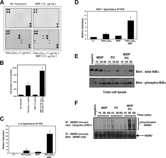
MDP, a NOD2 agonist, and PC, a TLR2 agonist, synergize to increase cytokine release, and this correlates with a prolonged NF-κB activation and with prolonged NEMO ubiquitination. (A) MDP and PC synergize to increase specific cytokine expression. THP-1 cells were either left untreated or were treated with 10 μg/ml MDP, 1 μg/ml PC, or 10 μg/ml MDP plus 1 μg/ml PC overnight. The medium was collected and subjected to cytokine array analysis (Ray Biotech, Inc.). The position of each of the 40 cytokines on the array is indicated in Fig. S3 in the supplemental material. In this analysis, there was limited cytokine release in either the untreated or the MDP-treated THP-1 cells (upper two panels). PC caused strong upregulation of IL-8 and RANTES and weaker upregulation of GRO (macrophage inhibitory protein β/γ [MIPβ/γ]) (lower left panel). Cells treated with MDP plus PC showed strong upregulation of IL-8, RANTES, MCP-1, IL-6, and GRO (MIPβ/γ) and weaker upregulation of TNF-α (lower right panel). (B) IL-6 ELISA performed under identical conditions shows an additive effect of PC and MDP. (C and D) Quantitative TaqMan RT-PCR was performed on THP-1 cells 2 h after stimulation with 500 ng/ml PC, 1 μg/ml MDP, or 500 ng/ml PC plus 1 μg/ml MDP. Total RNA from each sample was extracted and equalized. Within the RT-PCR cycle, each sample was then internally standardized to the 18S RNA present in that sample. Relative expression levels (with standard errors of the mean) are presented. (E) Macrophages were treated with PC (500 ng/ml), 10 μg/ml MDP, or both PC and MDP for 15, 30, or 60 min. Lysates were generated, and Western blots were performed. Phospho-IκB is shown in the lower blot. This blot was stripped and reprobed for total IκB (upper blot). (F) To correlate the synergy seen at the cytokine level with NEMO ubiquitination, macrophages were treated with 10 μg/ml MDP, 200 ng/ml PC, or both for 15, 30, or 60 min. In addition, one plate of cells was left untreated. At the indicated time, NEMO was immunopurified under stringent washing conditions and Western blotting was performed. MDP caused a slower and more prolonged NEMO ubiquitination, while PC caused a stronger, more acute NEMO ubiquitination (upper blot). When both MDP and PC were added, the NEMO ubiquitination was both more pronounced and more prolonged.

