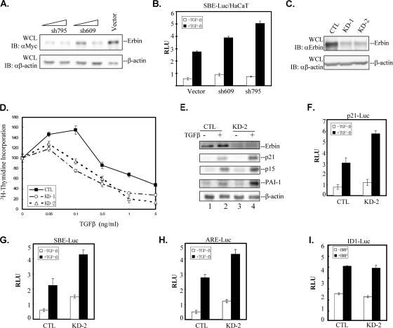
FIG. 6.
Knockdown of Erbin expression enhances TGFβ signaling. (A) Different shRNAs against Erbin reduce Erbin protein levels in HEK293T cells. Increasing amounts of Erbin shRNA construct shRNA-795 or shRNA-609 or the empty vector pSRG were transiently transfected into HEK293T cells together with Myc-Erbin. Knockdown of Erbin expression was examined by Western blotting. Immunoblotting with anti-β-actin antibody (αβ-actin) served as a loading control. (B) Reducing Erbin expression enhances TGFβ-induced SBE-Luc expression. HaCaT cells were cotransfected with the SBE-Luc reporter and Erbin shRNA construct shRNA-795 (sh795) or shRNA-609 (sh609). Luciferase activity was measured 20 h after TGFβ stimulation. (C) Erbin expression is effectively reduced in Erbin shRNA stable cell lines. Whole-cell lysates (WCL) were prepared from two independent HaCaT cell lines stably expressing Erbin shRNA-795 (KD-1 and KD-2) or an empty pSRG vector (CTL). The endogenous Erbin expression level was detected by Western blotting. Immunoblotting (IB) with anti-β-actin antibody served as a loading control. (D) Reducing Erbin expression enhances TGFβ-induced growth inhibition. KD-1, KD-2, or CTL cells were stimulated with various concentrations of TGFβ as indicated. Cell proliferation was assessed by incorporation of [3H]thymidine. Data are expressed as mean percentages ± standard deviations of thymidine incorporation relative to basal counts of each cell line from duplicate experiments. (E) Knockdown of endogenous Erbin expression enhances ligand-induced expression of TGFβ target genes. Whole-cell extracts from control HaCaT cells or KD-2 cells treated with TGFβ for 24 h were immunoblotted with the indicated antibodies shown on the right. The β-actin immunoblot served as a loading control. (F to H) Reducing Erbin expression enhances Smad2/Smad3-mediated transcriptional activity. HaCaT line KD-2 or CTL cells were transfected with p21-Luc (F), SBE-Luc (G), or ARE-Luc (H). Luciferase activity was measured 20 h after TGFβ stimulation. (I) Reducing Erbin expression did not affect BMP signaling. HaCaT line KD-2 or CTL cells were transfected with Id1-Luc. Luciferase activity was measured 20 h after BMP2 stimulation. RLU, relative luciferase activity; αMyc, anti-Myc antibody; αErbin, anti-Erbin antibody.

