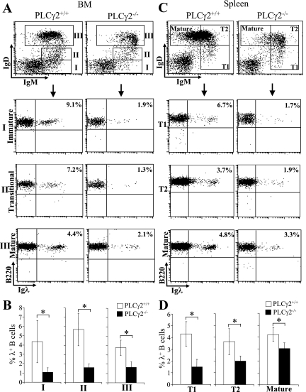
FIG. 2.
Reduction of Igλ chain expression during B-cell development in PLCγ2-deficient mice. (A) BM cells were isolated from wild-type (PLCγ2+/+) and PLCγ2-deficient (PLCγ2−/−) mice and stained with a combination of anti-B220, anti-IgM, anti-IgD, and anti-Igλ antibodies. Based on expression of IgM and IgD, late BM B cells were defined as IgM+ IgD− immature B-cell (I), IgM+ IgDlow transitional B-cell (II), and IgM+ IgD+ mature B-cell (III) subpopulations (top). The expression of B220 and Igλ in the gated B-cell subpopulations (I, II, and III) was analyzed (bottom). Percentages indicate Igλ+ B cells in the gated B-cell subpopulation. Data are representative of eight mice per genotype. (B) Statistical analysis of the percentages of Igλ+ B cells in the gated B-cell subpopulation from panel A (n = 8; *, P < 0.01). (C) Splenocytes were isolated from wild-type and PLCγ2-deficient mice and stained with a combination of anti-B220, anti-IgM, anti-IgD, and anti-Igλ antibodies. Based on expression of IgM and IgD, splenic B cells were defined as IgMhi IgD− T1 B-cell (I), IgMhi IgD+ T2 B-cell (II), and IgMlo IgD+ mature B-cell (III) subpopulations (top). The expression of B220 and Igλ in the gated B-cell subpopulations (I, II, and III) was analyzed (bottom). Percentages indicate Igλ+ B cells in the gated B-cell subpopulation. Data are representative of eight mice per genotype. (D) Statistical analysis of the percentages of Igλ+ B cells in the gated B-cell subpopulation from panel C (n = 8; *, P < 0.01).

