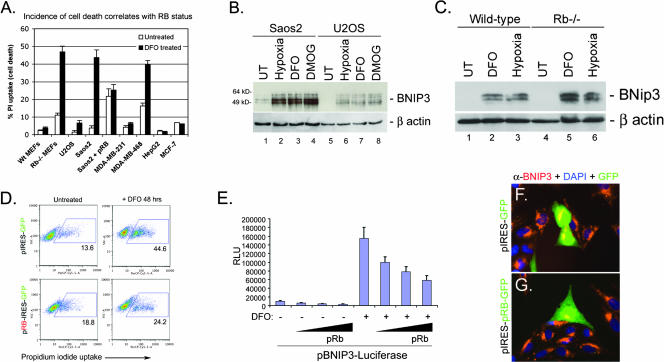
FIG. 4.
RB status and BNIP3 expression correlate with cell death. (A) The incidence of cell death of primary MEFs and different human tumor cell lines was assessed after 48 h of treatment with DFO by measuring uptake of PI, which was taken up only by dying cells. Wt, wild type. (B) Western blot analysis of BNIP3 induction in Saos2 and U2OS cells by hypoxia, DMOG treatment, or DFO treatment. (C) Western blot analysis of BNip3 induction in wild-type and Rb null MEFs by DFO treatment or hypoxia. UT, untreated. (D) Saos2 cells were transiently transfected with pIRES-GFP or pRB-IRES-GFP and treated with DFO for 48 h. The incidence of cell death was determined by flow cytometric analysis of PI uptake. (E) The effect of pRB overexpression for BNIP3 promoter activity was determined for Saos2 cells by a luciferase assay for reporter gene expression. Results presented are the averages of at least three independent experiments. RLU, relative luciferase units. (F and G) The effect of pRB expression on the levels of BNIP3 protein was determined by immunofluorescent staining for BNIP3 (red) in cells that expressed GFP by virtue of transfection with either pIRES-GFP (F) or pRB-IRES-GFP (G). DAPI, 4′,6′-diamidino-2-phenylindole.

