FIG. 7.
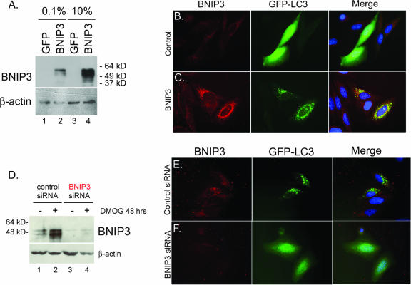
BNIP3 is required for hypoxia-induced autophagy. (A) Western blot analysis of BNIP3 in lysates from Saos2 cells that had been transfected with the control plasmid (pIRES-GFP) or with the BNIP3 expression plasmid (pBNIP3-IRES-GFP) under conditions of low-concentration (0.1%) or high-concentration (10%) serum. (B and C) The effect of BNIP3 overexpression (C, red) for the incidence of punctate GFP-LC3 staining (green) was assessed in Saos2 cells (grown in low-concentration serum) and compared to that observed in control cultures transfected with empty vector (B). (D) Western blot analysis of BNIP3 in lysates from Saos2 cells that had been transfected with either control siRNA or BNIP3 siRNA from untreated cells or cells treated with DMOG for 48 h. (E and F) Knockdown of BNIP3 (F, red) in hypoxic, serum-starved Saos2 cells blocked autophagy, as determined by loss of punctate GFP-LC3 staining (green), whereas no effect was observed with control siRNA.

