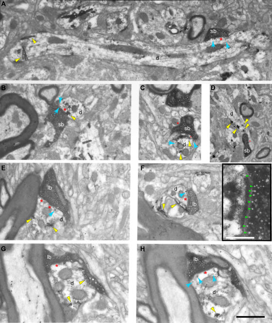
Figure 4. The fine structure of large and small prefrontal terminations in the ventral anterior nucleus.
The photographs were obtained from single two-dimensional sections. A–C, Electron microscopic (EM) photomicrographs showing small labeled boutons (sb) forming synapses with PV+ dendrites (d; black rods or grains, yellow arrowheads), with metabotropic mGluR1a receptors perisynaptically (black dots, blue arrows). D, EM photomicrograph showing a small labeled bouton forming a synapse with a PV+ dendrite (d; yellow arrowheads) without any mGluR1a receptors perisynaptically. E–H, EM photomicrographs showing large labeled boutons (lb) forming synapses with CB+ dendrites (d; black rods or grains, yellow arrowheads), with ionotropic NR1 receptors perisynaptically (black dots, blue arrows). The inset in F shows the synapse of that bouton at higher magnification showing docked vesicles (green arrowheads); scale bar: 0.25 µm. G and H show two serial sections of the same large bouton. The TMB labeling of the CB+ dendrite is obvious in G (yellow arrowheads), and the gold labeling of the perisynaptic NR1 receptors is clearly apparent in H (black dots and blue arrows). In many cases labeled receptor molecules were observed presynaptically (e.g., E) or inside axons (e.g., G). Red asterisks (*) indicate postsynaptic densities. Scale bar: 1 µm.

