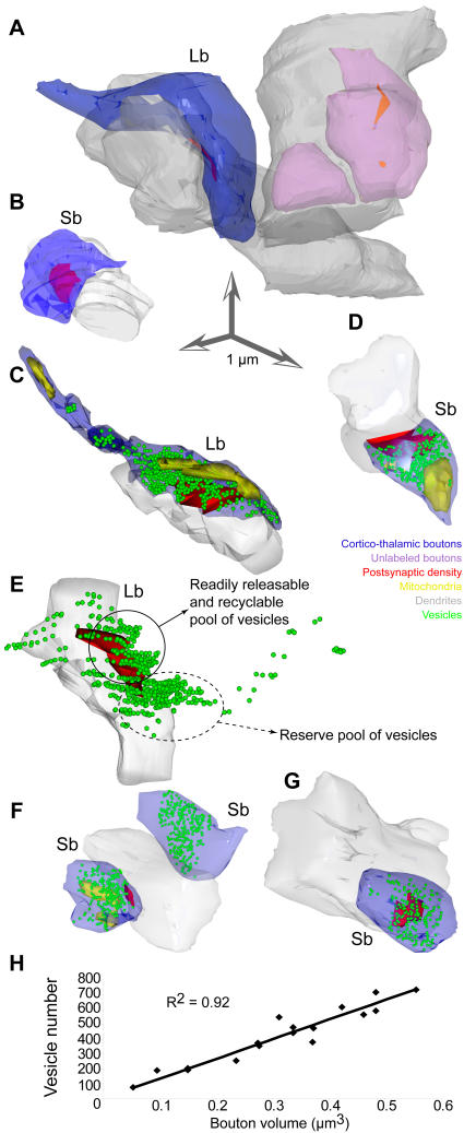
Figure 5. Presynaptic features of large (Lb) and small (Sb) boutons in the ventral anterior nucleus.
A-B, 3D reconstruction of labeled prefrontal boutons (blue; same as in Fig. 4B, G) forming synapses (red) with PV+ or CB+ dendrites (grey). Unlabeled boutons (pink) were included for comparison. C–G, The position of vesicles (green spheres) and mitochondria (yellow structures inside boutons) are shown in the reconstructions. The large bouton in C is a reconstruction of the bouton in Fig. 4E, and the small bouton in D is a reconstruction of the bouton in Fig. 4C. E, In most cases with large boutons (Lb), there were two separate clusters of vesicles: a smaller pool (solid circle), adjacent to the postsynaptic density (red), and a larger pool distal to the postsynaptic specialization (dotted circle). E, contains the same structures as C, but is rotated 180o in the y axis, and the bouton and its mitochondria were removed to facilitate distinction of vesicle clusters. Large boutons (Lb) were at least 2 times bigger than small boutons (Sb). H, The relationship of bouton volume to vesicular content was linear.

