
Figure 3.
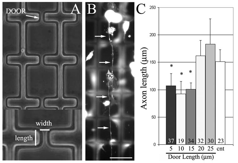
Axon length can be controlled using 3-D constraints. (A) Example of corridors with doors. Doors are described by their width and length. (B) Example of neuron extending axon (arrows) through multiple doors in a corridor. Some non-neuronal cells, fibroblasts, from the DRG are also present (labeled, nc). Focus is on the ‘floor’ of the corridor and walls are out of focus. Bar = 40 μm. (C) Graph of axon length as a function of door length. Doors of lengths less than 20 μm resulted in shorter axons than controls extending in corridors without doors (cnt). Numbers of axons analyzed per group is shown in the bars. Cells were immunocytochemically stained with anti-tubulin antibodies.