
Fig. 4.
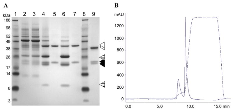
Purification of CPMV-PA1L VLPs. (A) SDS-PAGE analysis. The samples were run on 10% SDS-PAGE gel stained with Simply Blue Safe Stain (Invitrogen, San Diego, CA). Samples taken after the following purification steps were analyzed: Tissue disruption and clarification (lane 2), inactivation (lane 3), PEG precipitation (lane 4), anion exchange chromatography (lane 5 and 6), buffer exchange and sterile filtration (lane 7). Lanes 5 and 6 represent the first and second eluted peak, respectively. Lane 1 and 8 is the SeeBlue Plus2 molecular weight marker (Invitrogen, San Diego, CA). Lane 9 is the purified wild type CPMV control. The uncleaved CPMV-PA1L L coat protein and wild type CPMV L coat protein are indicated by open triangles. The full-length and trimmed S coat proteins are indicated by closed triangles. The CPMV-PA1L L coat protein cleavage products are indicated by dashed triangles. (B) CPMV-PA1L anion exchange chromatogram. Absorbance at 260 nm is indicated on the Y-axis and time in minutes is indicated on the X-axis. The conductivity trace is shown (dashed line).