
Fig. 1.
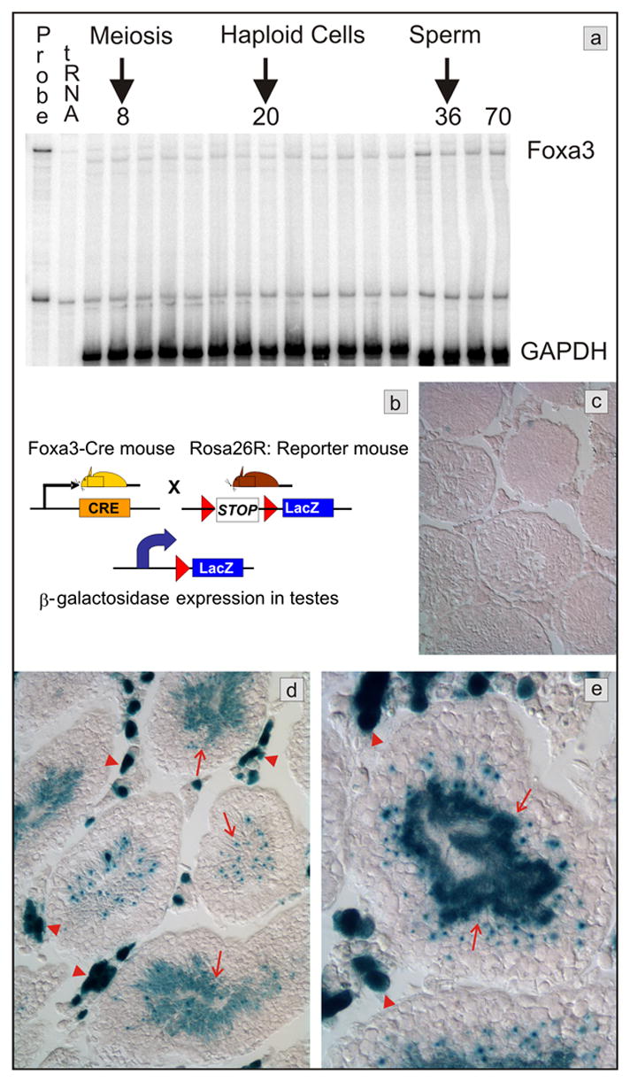
Testicular expression of Foxa3. a) Messenger RNA expression analysis by RNase protection assay. Foxa3 mRNA is detectable in the mouse testis throughout postnatal development. The mRNA abundance is increased approximately 2-fold in the adult testis when compared to the 6 days old testis (normalized to GAPDH mRNA). RNA was prepared from testis of postnatal day 6, 8, 10, 12, 14, 16, 18, 20, 22, 24, 26, 28, 30, 32, 36, 40, and 70, respectively. b) Scheme of the genetic lineage tracing employed to track Foxa3 expression in the testis. Beta-galactosidase expression occurs only in those cells that express Foxa3 or are the descendants of Foxa3-expressing cells. c) The control mouse (Rosa26R, no Cre), shows no β-galactosidase expression in the testes. d, e) Mice carrying both the β-galactosidase reporter and the Foxa3-Cre transgene (Rosa26R; Foxa3Cre mice) display robust staining in Leydig cells (red arrowheads) and in spermatids (arrows).