
Fig. 2.
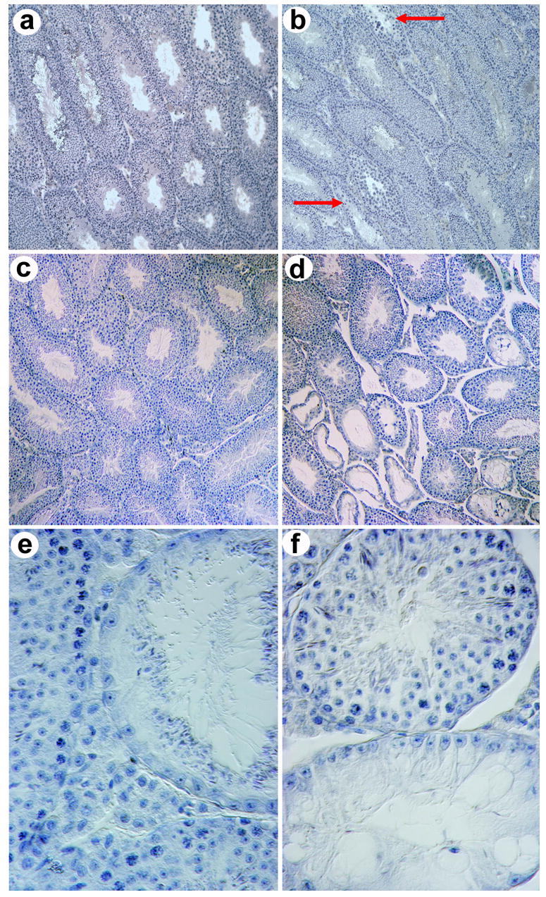
Testicular degeneration in Foxa3-deficient mouse testes. a) Histological section of a wild type mouse testis at the age of three months. All seminiferous tubules contain somatic Sertoli cells as well as germ cells and exhibit ongoing germ cell production. b) Mutant testis at the age of three months. Sporadic tubulus with early signs of degeneration, i.e. thinning of the germinal epithelium, can be seen (arrow), c) Histological section of a wild type mouse testis at the age of eight months showing normal spermatogenesis in all tubules. d,e,f) Histological sections of Foxa3-deficient mouse testes at eight months of age. The testis contains both tubules with normal appearance and ongoing spermatogenesis as well as severely degenerated tubules. The characteristic feature of the defective tubules is a selective loss of germ cells. The Sertoli cells remained within the tubule resulting in a focal Sertoli-cell-only syndrome. e) An almost completely atrophied seminiferous tubule. The only germ cells present are elongated spermatids. All spermatogonia, spermatocytes, and round spermatids are missing. The elongated spermatids are arrayed on the adluminal surface of the Sertoli cells as they usually are shortly before sperm release from the germinal epithelium (spermiation). f) A Sertoli cell only tubule (bottom) adjacent to a tubule with complete spermatogenesis (top). The Sertoli cell nuclei characterized by their triangular shape and their prominent nucleoli have aligned with the basal lamina indicating the absence of any germ cells. Heterozygous testes show histological pictures similar to the homozygous mutant testes. Magnification of a), b), c), and d) 100x; e) and f) 600x.