
Fig. 3.
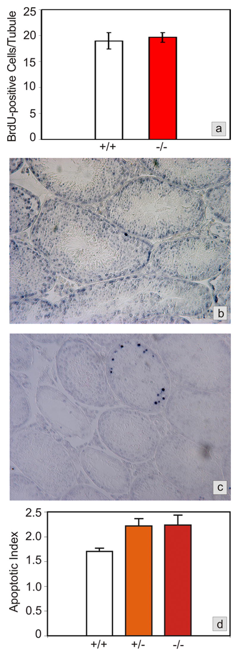
Testicular proliferation and apoptosis in Foxa3-deficient mice. a) Proliferation rates, as determined by BrdU labeling of cells in S-phase during spermatogenic stages VII and VIII, are not different between wild type and Foxa3-deficient mice (p>0.35). b) Apoptosis in a wild type mouse testis as revealed by the TUNEL-technique. Only a minor proportion of all tubules contains one or more TUNEL-positive cells. All other tubules are devoid of TUNEL-positive cells. As judged from the appearance and the location of the cells in the seminiferous tubule the apoptotic cells are almost exclusively spermatogonia and spermatocytes. c) Foxa3-deficient mice loose their germ cell population by apoptosis. The tubule undergoing degeneration exhibits a high number of apoptotic germ cells, again mainly spermatogonia and spermatocytes. Note the already degenerated tubules which are devoid of TUNEL-positive cells. d) Apoptotic index expressed as TUNEL-positive cells per tubule containing TUNEL-positive cells. The mutant as well as the heterozygous testes show a significant increase in the number of apoptotic cells (p<0.05).